
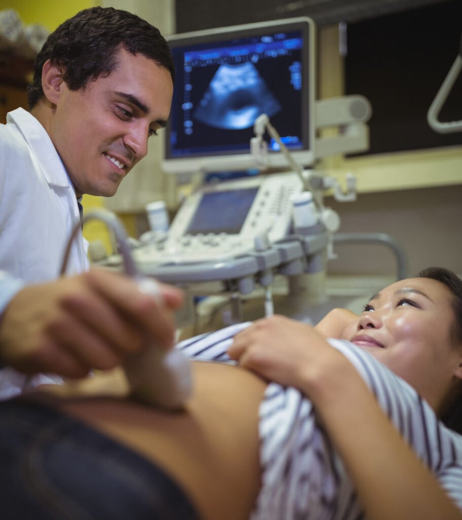
 Ultrasound (US) offers a myriad of benefits in the field of medical diagnostics and imaging. One of its primary advantages is its non-invasive nature, as it does not involve the use of ionizing radiation, making it a safe and widely accessible tool for various patient populations, including pregnant women and children. Ultrasound is especially valuable in obstetrics, providing detailed images of the developing fetus during pregnancy, enabling healthcare professionals to monitor fetal growth, detect anomalies, and assess overall well-being. Additionally, ultrasound plays a crucial role in cardiovascular imaging, allowing for the visualization of the heart’s structure and function, aiding in the diagnosis of conditions such as heart valve abnormalities and congenital heart defects.
Moreover, ultrasound is an essential tool in the examination of abdominal organs, such as the liver, kidneys, and gallbladder, facilitating the detection of tumors, cysts, and other abnormalities. Its real-time imaging capability makes it valuable in guiding medical procedures, such as biopsies or needle aspirations, ensuring precision and accuracy. Furthermore, ultrasound has become an integral part of musculoskeletal imaging, aiding in the diagnosis and monitoring of conditions affecting the joints, muscles, and tendons.